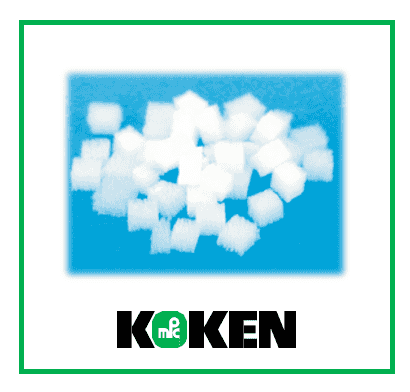
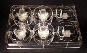
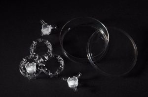
The ‘Honeycomb’ collagen sponge has a structure in which uniform pores (250-400 um) are arranged densely in one direction, into which cells can penetrate and proliferate. This structure facilitates the ready supply of nutrients to the cells inside the sponge, and releases metabolic wastes and biochemical products. Cells can proliferate and fill the lumen to form a uniform cell mass. The ‘Honeycomb’ collagen sponge is prepared from highly purified bovine dermal type I Atelocollagen and can be degraded by collagenase.

Suppression of tumorigenesis from transplanted mouse ES cells by cultivation with Atelocollagen Honeycomb Sponge
(M. Yamaki, Matsumoto Dental University, Japan)
Mouse ES cells cultured on honeycomb sponge were transplanted by subcutaneous injection into 6-week-old mice. Although all embryoid bodies without the honeycomb sponge formed tumor without exception, none of the embryoid bodies transplanted with the honeycomb sponge formed tumors (Fig.1).
Histological analysis of transplanted (mouse ES/+CSH) and normal kidney showed no differences between the two tissues. The results indicates normal spontaneous differentiation of the mouse ES cells in the presence of the honeycomb sponge when transplanted in vivo (Fig.2).

SPECIFICATION
Product Name
Atelocollagen Honeycomb Sponge
Catalog Number
KKN-CSH-10
Size
100 mg (bottle)
Sponge Dimensions
3 × 3 × 2 mm sponges
12-13 mg/mL density
200-400 µm pore size
approx. 2000 cm² / g surface area
Storage and Stability
Store at Room Temperature
Sterility
Sterile
Notice To Purchaser
REPROCELL is a licensed global distributor of KOKEN’s collagen-derived products everywhere, except for Japan.
Manufacturer
Koken Co., Ltd. (Japan)
Warning: call_user_func() expects parameter 1 to be a valid callback, function 'my_shipping_tab_callback' not found or invalid function name in /home/anhsci/domains/anhsci.com/public_html/wp-content/themes/flatsome/woocommerce/single-product/tabs/tabs.php on line 70

